The wonderful world of
Basidiobolus
Hello, and welcome to
my blog about this very interesting fungus; Basidiobolus. My name is
Michelle Austin, and I am currently working on my masters in biology at Western
Illinois University. My research focuses on Geomyces destructans and other keratinophilic fungi species but I have a passion for all fungi. I am currently taking medical mycology and have become quite
interested in fungal pathogens such as Basidiobolus. I hope you enjoy reading about this
fungus as much as I have.
General Description of
Basidiobolus
Taxonomic
Classification:
Kingdom: Fungi
Phylum: Zygomycota
Subphylum: Zygomycotina
Class: Zygomycetes
Order: Entomophthorales
Family: Basidiobolaceae
Genus: Basidiobolus
Phylum: Zygomycota
Subphylum: Zygomycotina
Class: Zygomycetes
Order: Entomophthorales
Family: Basidiobolaceae
Genus: Basidiobolus
Zygomycota fungi make up about 1% of the total number of fungi inhabiting the world yet these fungi are the primary colonizers of most substrates. Basidiobolus is a member of this phylum and is a filamentous fungus that
is most commonly found in soil, decaying organic matter, and the
gastrointestinal tracts of amphibians, reptiles, fish and bats. Basidiobolus is the sole genus of the family Basidiobolaceae which has classically been the second family recognized in the order Entomophthorales [12]. The two most common species are B. ranarum and B. haptosporus [12]. Although
this fungus can be found worldwide, human infections caused by this fungus are
most commonly reported from Africa, South America and tropical Asia [1]. This could be due to the fact that only pathogenic species of this fungus are found in those regions or maybe the environmental conditions in these areas are more favorable to the pathogenic species.
Basidiobolus ranarum is an an example of a pathogenic species in Basidiobolus fungi. This species is an opportunistic pathogen which means it occasionally causes disease in humans (immunocompromised individuals or young children). Since it is often found in soil and in vertebrate guts and dung especially of amphibians and reptiles [10], it would be very easy for children to come in contact with this fungus. Basidiobolus is dimorphic meaning it can grow as hyphae and/or yeast depending upon the environmental conditions. An important feature of hyphal growth of this species is the cytoplasm migrates with the growing tip. This growth causes mycelium that consists of mostly empty cells and a collection of individual growing tip cells with no internal sharing of resources or genetic material. One unique characteristic of Basidiobolus is that it has extremely large nuclei (25–50 µm) [10] compared to most filamentous fungi which have nuclei with a diameter of 1-3µm [11]. The nucleoli of Basidiobolus can fill nearly the entire nucleus [10]. *See diagram below*
 |
| Diagram of a fungal cell http://www.peteducation.com/article.cfm?c=16+2160&aid=2956 |
Rate
of Growth: Since this fungus is a Zygomycota fungus, growth is rapid and the fungus matures within 5 days. Maturity can be determined by whether or not the fungus is producing spores [1].
Colony
Morphology: Colonies of this fungus are flat, thin,
waxy in appearance and can vary in color buff to gray. Later growth of the fungus
can be characterized by its heaped up or radially folded appearance and grayish
brown color. This later growth of the fungus is covered by fine, white, powdery
surface mycelium. If you reverse the plate, the colony looks white or pale. An
important note: some strains have an earthy odor typical for Streptomyces spp (a bacteria) [2].
Culture of Basidiobolus ranarum
http://www.mycology.adelaide.edu.au/Fungal_Descriptions/Zygomycetes/Basidiobolus/ |
*Important note: Basidiobolus species lose their sporulation
ability after relatively short periods of time in culture. The use of media
which incorporates glucosamine hydrochloride and casein hydrolysate seems to
help overcome this problem [7]. The media also appears to be suitable for
culturing Conidiobolus species, which is interesting considering the two fungal
species are considered lookalikes.
Microscopic
features:
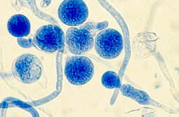
The hyphae of Basidiobolus are large (8 to
20 µm in diameter) and septate. Having large hyphae/thick fungal walls are a common characteristic of fungi in the phylum Zygomycota. The more spores the fungus produces, the
greater the number of septa are present. Basidiobolus spp. produce
sexual spores called zygospores that are thick-walled and can be smooth or have
undulating outer cell walls. This fungus can also produce two different types
of asexual spores or conidia called: ballistospores and capilliconidia [2].
|
 |
| Asexual
spores of Basidiobolus ranarum http://www.mycology.adelaide.edu.au/Fungal_Descriptions/Zygomycetes/Basidiobolus/ |
Definitions:
hyphae: the branching filaments of a fungus
mycelium: a collection of hyphae
zygospores:sexual spores of fungi within the Zygomycota phylum
septate fungi: fungal hyphae that have partitions (septations)
sporangiophore: asexual stalk which a sporangium is attached
sporangium: asexual structure that holds sporangiospores
sporangiospores: asexual spores
Life Cycle of
Zygomycota fungi
Life
Cycle of a typical Zygomycota fungus
http://micro-organic-world.blogspot.com/2009/11/identification-of-medically-important.html |
An interesting feature of the life cycle of Basidiobolus is that it has the ability to produce several types of spores; usually, depending on its microhabitat. The first type of spore are ballistospores which is forcibly discharged to stick to substrate. The second type of spore are capilliconidia which is not forcibly discharged, but it has a weak spot in the conidiophore so that upon contact the spore is released, and capilliconidium with a sticky blob on glue on the tube-like haptor at its distal (outward) end can attach to anything it contacts, often a passing insect or even a growing fungal hypha. These capilliconidia may develop directly from a hypha or from a mature ballistospore [8].
Clinical Cases
Pathogenicity: Basidiobolus ranarum is the causative agent of subcutaneous
zygomycosis, which is a chronic inflammatory or granulomatous disease. It is
generally restricted to the limbs, chest, back or buttocks. The lesions are
very large in size, hard, palpable, nonulcerating subcutaneous masses. This
pathogenic fungus can also cause gastrointestinal infections on occasion [3]
This disease occurs usually in children, occasionally in adolescents and rarely
in adults. It more frequently affects males than females [3].
Below are links to
clinical cases associated with this fungus:
Case 1: http://jcm.asm.org/content/39/6/2360.full
Case 2: http://www.ncbi.nlm.nih.gov/pmc/articles/PMC2893432/
Case 1: describes a 41 year old male with a gastrointestinal infection
caused by Basidiobolus ranarum. The patient was admitted to the hospital
after complaining of abdominal pain. An ultrasound revealed a thick mass in the
right iliac fossa and showed a marked thickening of the ascending colon and
cecum and an increased echogenicity of the renal cortex. His prostate was
enlarged and he also had a fecal impaction [4]. After an intestinal
biopsy, their original diagnosis was intestinal tuberculosis but when it was cultured,
there was no growth of mycobacterium. A tissue biopsy was not performed to
check for a fungal infection, however his urine cultures came back with fungal
growth positive for Basidiobolus ranarum. The patient was treated with
Amphotericin B, however his condition did not improve and they later realized
that his strain of B. ranarum was resistant to Amphotericin B. After 4
weeks, the patient decided to return home to his family. Since there have only
been 10 reported cases in literature, surgical resection of the infected tissue
and prolonged treatment with itraconazole appear to be the best available
options for treatment [4].
Case 2: describes a 42 year old diabetic male with
suspected lung abscesses and multiple organ damage. He is a smoker, works on a
farm, and has had diabetes since he was 20. He had generalized hyperkeratotic
skin lesions with central necrosis located on his lower limbs and back of
chest. Some of these lesions were simulating diabetic kyrle and some healed
pyoderma [5]. An examination of the respiratory system revealed a diagnosis of
non-resolving pneumonia right upper lobe. Since the X ray appearance was not
typical of a lung abscess, there were multiple possibilities for diagnosis such
as: fungal infection, mycobacterial infection or hydatid cyst. A CT of the
thorax revealed fluid and solid attenuation areas that were mimicking a fungal
ball as well as double layered cyst wall as in hydatid cyst. He was initially
treated with a broad spectrum of antibiotics and his condition did not improve.
Blood heart infusion broth revealed growth of B. ranarum and the patient was
started on treatment right away. His treatment consisted of potassium iodide
which was considered the drug of choice for this fungus. He was started on oral
potassium iodide with 1 drop 3 times a day and gradually increased to 10 drops
3 times a day. He was also given cotrimoxazole. Since the patient's condition
did not permit the use of the specific agent-KI, they had to perform surgery to
remove the fungus. He was taken to cardio-thoracic surgery for resection after
control of diabetes and renal function where a right upper lobectomy was done.
The patient completely recovered after this [5].
Below is an image of
the destruction caused by B. ranarum
 |
| Cutaneous
lesions caused by B. ranarum http://www.mold.ph/basidiobolus.htm |
Geographic
Distribution
Distribution: Basidiobolus is a ubiquitous fungus and the majority of cases have been reported in South
America, Africa and tropical Asia [1]. However, recently (2012) this fungus has
been classified as an emerging invasive fungal infection in desert regions of
the US Southwest causing gastrointestinal basidiobolomycosis [9].
 |
| Map
of the world showing areas where human infections are reported www.ezilon.com |
How do people acquire
this fungal infection? Traumatic
implantation or insect bite are the most likely way for B. ranarum to
enter the body [5].
Habitat: Basidiobolus can be found in the feces of amphibians, reptiles, and insectivorous bats as well as in wood lice, on decaying fruits and organic matter and soil [2].
Look Alikes: Conidiobolus is quite similar to Basidiobolus however, Conidiobolus have sporangiophores that are not swollen at their apices and zygospores that do not contain beaks [6].
|
 |
| Asexual
spores of Condiobolus http://www.mold.ph/conidiobolus.htm |
Websites you should
check out:
References
[1] Gugnani H.C.
1999. A review of zygomycosis due to Basidiobolus ranarum. European Journal
of Epidemiology 15: 923-929
[2] Rex J.H., McGinnis M., Arikan S., Rodriquez-Adrian L., Kirsch M. 2011 Basidiobolus Species www.drfungus.org/thefungi/Basidiobolus.php (March 29th 2012) [3] Larone D.H. 2002. Medically important fungi A guide to identification 4th edition. ASM Press page 176 [4] Kahn Z.U., Khoursheed M., Makar R., Al-Waheeb S., Al-Bader I., Al-Muzaini A., Chandy R., Mustafa A.S. 2001. Basidiobolus ranarum as an Etiologic Agent of Gastrointestinal Zygomycosis. Journal of Clinical Microbiology vol 39 no. 6: 2360-2363 [5] Chetambath R., Deepa Sarma M.S., Suraj K.P., Jyothi E., Mohammed S., Philomina B.J., Ramadevi S. 2010. Basidiobolus: An unusual case of lung abscess. Lung India 27(2): 89-92 [6] Rex J.H., McGinnis M., Arikan S., Rodriquez-Adrian L., Kirsch M. 2011 Conidiobolus Species http://www.doctorfungus.org/thefungi/Conidiobolus.php (March 29th 2012)]
[7] Shipton W.A.,
Zahari P., 1987 Sporulation Media for Basidiobolus species Journal Med Vet
Mycology 25(5):323-327
[8] Blackwell M.,
2001 Fungi that fly: A two part laboratory study Mycology laboratory manual:
LSU
[9] Vikram H.R., Smilack J.D., Leighton J.A., Crowell M.D., De Petris G. 2012 Emergence of
Gastrointestinal Basidiobolomycosis in the United States, With a Review of Worldwide Cases Clinical Infectious Diseases
[10] Henk D.A., Fisher M.C., 2012The Gut Fungus Basidiobolus ranarum Has a Large Genome and Different Copy Numbers of Putatively Functionally Redundant Elongation Factor Genes PLoS ONE 7(2): e31268. doi:10.1371/journal.pone.0031268
[11] Gow N.A., Gadd G.M., 1994 The Growing Fungus. Chapman & Hall pgs: 75-76
[12] Howard D.H., 1983 Pathogenic Fungi in Humans and Animals. Library of Congress pgs 130-132 |


No comments:
Post a Comment